Scientists have developed cutting-edge MRI technology that can diagnose aortic stenosis more quickly and accurately than traditional methods, marking a major advance in cardiac imaging.
Aortic Stenosis: A Widespread and Dangerous Condition
Aortic stenosis, a serious and progressive heart condition, affects approximately 300,000 people in the UK and about 5% of Americans aged 65 and older. The disease occurs when the aortic valve—the heart’s main outflow valve—narrows and stiffens, restricting blood flow from the heart to the body. Common symptoms include chest pain, palpitations, dizziness, shortness of breath, and fatigue, especially during physical activity.
New Study Confirms Superior Accuracy of 4D Flow MRI
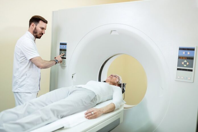
In a recent study, researchers from the University of Sheffield and the University of East Anglia (UEA) investigated the effectiveness of a four-dimensional flow (4D flow) MRI scan in diagnosing aortic stenosis. Unlike conventional ultrasound (echocardiography), which can sometimes underestimate disease severity, the 4D flow MRI delivers more accurate and reliable measurements of blood flow through heart valves.
Improved Decision-Making and Earlier Interventions
Professor Andy Swift, from the University of Sheffield’s School of Medicine and Population Health and an Honorary Consultant Radiologist, emphasized the clinical impact of the new technology:
“4D flow scanning holds significant promise to improve how we assess aortic stenosis. Its enhanced accuracy may allow for earlier and more precise diagnosis. Better measurements help clinicians determine exactly when surgical intervention is needed, potentially reducing complications.”
A Closer Look at Blood Flow in Time and Space
Dr. Pankaj Garg, lead researcher from UEA’s Norwich Medical School and Consultant Cardiologist at Norfolk and Norwich University Hospital, explained the innovation:
“Unlike ultrasound, 4D flow MRI lets us see blood flow in three spatial directions over time, the fourth dimension. This level of detail could transform how we detect and treat aortic stenosis.”
He noted that current ultrasound techniques, while widely used, sometimes fail to capture the full extent of disease, which can delay life-saving treatments.
Clinical Validation Through Patient Comparison
To validate the technology, the research team examined 30 patients previously diagnosed with aortic stenosis. Each patient underwent both traditional echocardiography and 4D flow MRI scans. By comparing the results, researchers assessed which imaging technique more accurately identified patients needing timely valve intervention.
They then tracked the patients’ clinical outcomes over eight months to confirm their findings.
Results Demonstrate Clear Superiority of 4D Flow MRI
As reported by sheffield.ac.uk, the study, which included key contributions from Dr. Samer Alabed and Professor Andy Swift, found that 4D flow MRI consistently outperformed traditional ultrasound in assessing the severity of aortic stenosis. The new technology delivered more reliable data, helping doctors make better-informed treatment decisions.
A Potential Lifesaver for Thousands
With its improved diagnostic capabilities, 4D flow MRI could save thousands of lives, especially by enabling earlier interventions and reducing complications associated with delayed treatment.


