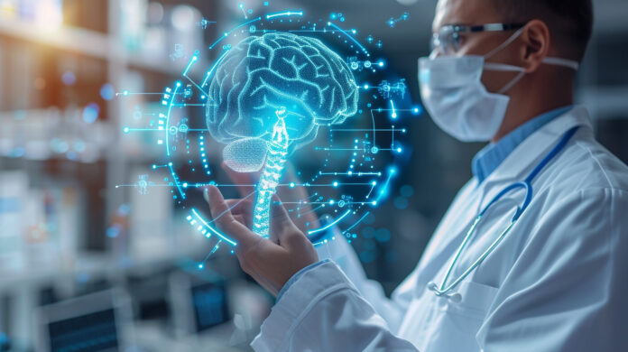
A recent study by Karl Landsteiner University of Health Sciences (KL Krems) demonstrated the effectiveness of machine learning (ML) in swiftly and accurately diagnosing mutations in gliomas and primary brain tumors. This study utilized physio-metabolic magnetic resonance imaging (MRI) data analysis to identify mutations in a metabolic gene crucial for disease prognosis and treatment planning.
Gliomas pose significant challenges due to their location within the brain, hindering easy access to individual tumor data necessary for personalized therapies. However, imaging techniques like MRI offer valuable insights, albeit with complexities in analysis. To address this, the Central Institute for Medical Radiology Diagnostics at St. Pölten University Hospital implemented ML and deep learning methods to automate and streamline such analyses for clinical integration.
One key mutation identified in the study was in the isocitrate dehydrogenase (IDH) gene, influencing clinical outcomes. Utilizing ML, the team achieved a remarkable precision of 91.7% and an accuracy of 87.5% in distinguishing between wild-type and mutated tumors. However, challenges arose from inconsistencies in data collection protocols across hospitals, impacting ML analyses’ effectiveness.
Despite standardization challenges, ML-based evaluation of physio-metabolic MRI data proved promising for preoperative determination of IDH mutation status, facilitating tailored treatment strategies for glioma patients.


