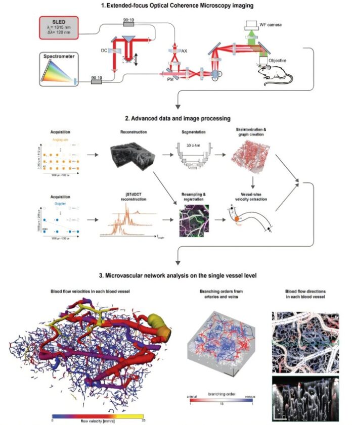

Researchers have developed a groundbreaking method using a Bessel beam to enhance the focus of optical coherence microscopy, enabling detailed imaging of large brain sections. This innovation bridges the gap between traditional methods, which either focus on small volumes or sacrifice detail over broader areas, by offering a comprehensive view of the brain’s vascular network.
As reported by medicalxpress, the technique combines high-sensitivity Doppler optical coherence tomography with advanced data processing algorithms to measure blood flow velocities and directions across thousands of brain vessels. This enables researchers to examine the relationship between blood flow, vessel size, and connectivity within the brain’s intricate microvascular system.
Understanding the vascular network is essential for studying brain function and neurological diseases like Alzheimer’s and stroke. By allowing researchers to observe changes in blood flow and vessel connectivity over time, this method provides valuable insights into the development and progression of such conditions. The findings were published in *Light: Science & Applications*.
A key advantage of this approach is its ability to visualize these changes in a living brain, making it possible to assess how potential treatments impact brain blood flow. This represents a significant leap forward in neurological research.
The team plans to extend the technology to study neurovascular conditions, starting with stroke and its treatments. They also envision broader applications, including research on other organs and diseases, which could pave the way for targeted vascular therapies.


