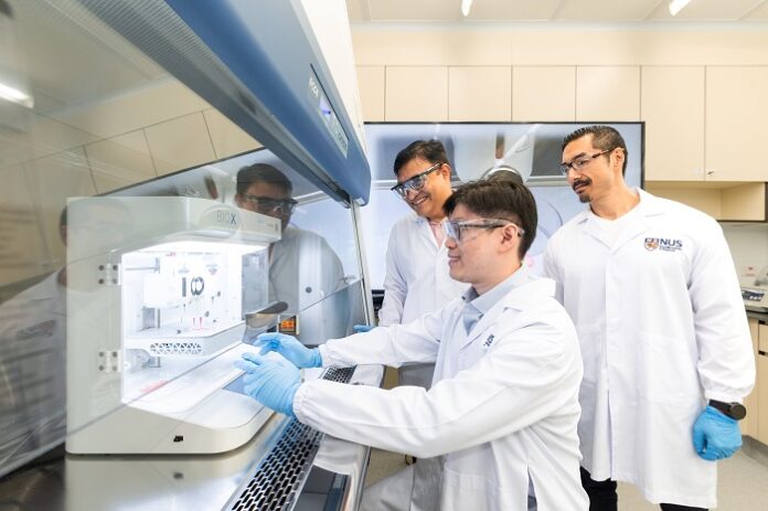

A team of researchers from the National University of Singapore (NUS) has developed a breakthrough method to fabricate customized gingival (gum) tissue grafts. By combining 3D bioprinting with artificial intelligence (AI), the team offers a less invasive and highly personalized alternative to traditional gum grafting procedures.
Led by Assistant Professor Gopu Sriram from the NUS Faculty of Dentistry, this approach eliminates the need to harvest tissue from a patient’s mouth—an uncomfortable process often limited by the availability of healthy tissue.
Addressing Critical Challenges in Gum Repair
Gum tissue grafts are crucial for treating mucogingival defects such as gum recession or damage from periodontal disease and dental implants. Traditionally, dentists harvest grafts from the patient’s own mouth. While effective, this method often leads to patient discomfort, limited tissue availability, and a higher risk of postoperative complications.
To solve these issues, the NUS team developed a 3D bioprinting method that fabricates soft tissue constructs tailored to the exact dimensions of a patient’s gum defects. Their bio-ink formulation supports healthy cell growth and ensures accurate printability, enabling the creation of stable, biomimetic grafts.
Accelerating the Process with Artificial Intelligence
Fine-tuning the 3D bioprinting process has traditionally required labor-intensive trial and error. Parameters like extrusion pressure, nozzle diameter, bio-ink viscosity, and printhead temperature heavily influence the outcome. This complexity often demands thousands of experiments to optimize results.
To streamline the process, the team integrated AI into their workflow. According to Professor Dean Ho, Head of Biomedical Engineering at NUS and co-author of the study, the AI algorithm reduced the number of necessary experiments from thousands to just 25 combinations, drastically cutting time and resource use.
“This approach allows us to fabricate high-quality tissue constructs with greater speed and precision,” Prof. Ho said. “It also ensures the structural integrity and cell viability of the final grafts.”
Achieving High-Quality, Biomimetic Gum Tissue
The bioprinted grafts demonstrated over 90% cell viability immediately after printing and maintained their structural properties during an 18-day culture period. Histological analysis confirmed the presence of key proteins and a multi-layered structure that closely mimics natural gum tissue.
“This is one of the first studies to directly combine 3D bioprinting and AI for oral soft tissue biofabrication,” said Asst Prof Sriram. “Because we’re working with living cells, bioprinting is much more complex than traditional 3D printing.”
A Transformative Future for Regenerative Dentistry
This innovation could transform dental care by enabling efficient, personalized tissue grafting for patients with periodontal disease or dental implant complications. By producing custom-fitted grafts, dentists can minimize invasiveness, shorten surgery time, and enhance recovery outcomes.
“This research shows how AI and precision medicine can converge to address longstanding clinical problems,” said Asst Prof Sriram. “Custom gum grafts can reduce tissue tension during wound closure and improve healing while minimizing discomfort.”
Beyond Dentistry: Implications for Other Tissues
According to Dr. Jacob Chew, a periodontist and co-investigator, this technique may also benefit other barrier tissues, like skin. Since oral tissues exhibit scarless healing, the approach could guide the development of skin grafts that promote scar-free wound recovery.
As reported by medicalxpress, the team plans to advance their findings through in vivo studies, exploring graft stability and integration in real oral environments. They also aim to introduce blood vessel integration through multi-material bioprinting. This advancement could enable more complex and functional tissue constructs.
Paving the Way for Broader Applications in Tissue Engineering
With these next steps, the researchers hope to revolutionize regenerative dentistry and expand the boundaries of tissue engineering. Their method holds immense promise not only for dental patients but also for broader clinical applications in regenerative medicine.


