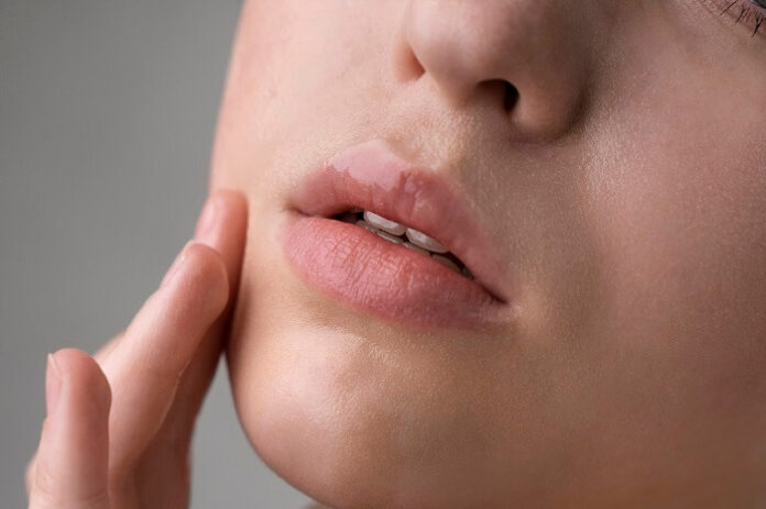
Our lips play vital roles in speaking, eating, drinking, breathing, and expressing emotions, as well as signaling health and beauty. Given their complexity, effective lip repair can be challenging. Basic research is key to advancing treatments, but until now, no specific models using lip cells—which function differently from other skin cells—have been available.
In a recent study, researchers have successfully immortalized donated lip cells, paving the way for the development of clinically relevant lip models in laboratories. This breakthrough, once scaled up, could significantly benefit many patients.
“The lip is a highly visible facial feature,” said Dr. Martin Degen of the University of Bern. “Defects in this tissue can be very disfiguring. However, we lacked human lip cell models to help create treatments. Our collaboration with the University Clinic for Pediatric Surgery and Bern University Hospital enabled us to change this by using lip tissue that would have otherwise been discarded.”
Primary cells directly donated from individuals are ideal for such research, as they closely resemble the original tissue. However, these cells can’t be reproduced indefinitely and are often hard to source.
“Human lip tissue is rarely obtainable,” explained Dr. Degen. “Without these cells, accurately mimicking the lip’s characteristics in vitro isn’t feasible.”
To overcome this, the team aimed to create immortalized lip cells that could be grown in the lab. They did so by altering specific gene expressions to allow continuous cell reproduction, beyond the normal cellular life span.
The scientists used skin cells from tissue donated by two patients, one with a lip laceration and another with a cleft lip. They employed a retroviral vector to deactivate a gene that halts the cell cycle and lengthened the telomeres on each chromosome, increasing cell longevity.
They rigorously tested the new cell lines to confirm genetic stability during replication and ensure they retained the properties of primary cells.
To rule out cancer-like changes, they checked for chromosomal abnormalities and attempted to grow the cells on soft agar, a medium only cancer cells can thrive on. The new cell lines showed no abnormalities and couldn’t grow on the agar, behaving similarly to primary cells in protein and mRNA production.
As reported by medicalxpress, to assess the cells’ suitability for future models of lip healing or infection, the researchers conducted additional tests. When scratched, untreated cells closed the gap within eight hours, while growth factor-treated cells healed faster—results consistent with wound healing in skin cells from other areas.
Next, the scientists created 3D models using the cells and infected them with *Candida albicans*, a yeast that can seriously affect people with weak immune systems or cleft lips. The pathogen successfully invaded the model, mirroring real lip tissue infection.
“Our lab is focused on understanding the genetic and cellular pathways involved in cleft lip and palate,” said Dr. Degen. “Yet, we believe 3D models of healthy immortalized lip cells have potential across various medical fields.”
He added, “One challenge is that lip keratinocytes may be of labial skin, mucosal, or mixed types. Depending on the research focus, specific cell types might be needed, but we have the tools to characterize and purify these populations in vitro.”


