


Researchers at University College London (UCL) have developed a groundbreaking handheld scanner that can produce detailed 3D photoacoustic images in just seconds, potentially transforming clinical diagnostics. This innovative device, outlined in a study published in Nature Biomedical Engineering, allows doctors to receive real-time scans using photoacoustic tomography (PAT), providing clear and accurate images of blood vessels. The technology could enable earlier diagnosis of conditions like cancer, cardiovascular disease, and arthritis.
PAT imaging utilizes laser-generated ultrasound waves to capture subtle changes in small blood vessels, offering a key early marker of disease. However, previous scanners were too slow for clinical use, requiring patients to remain completely still for over five minutes, which often led to blurry, unusable images. The new UCL-developed scanner speeds up the process, capturing high-quality 3D images in seconds, making it more practical for clinical settings.
Professor Paul Beard, who led the study, noted that this technology is 100 to 1,000 times faster than previous models, allowing real-time imaging of physiological events and reducing the impact of patient movement during scans. “These advancements make the system suitable for clinical use for the first time, allowing us to explore aspects of human biology and disease that were previously inaccessible,” he explained.
As reported by medicalxpress, the new scanner could revolutionize the diagnosis of inflammatory arthritis, which requires scanning all 20 finger joints in both hands. The new device can perform this scan in just a few minutes, a significant improvement over older scanners that took almost an hour.
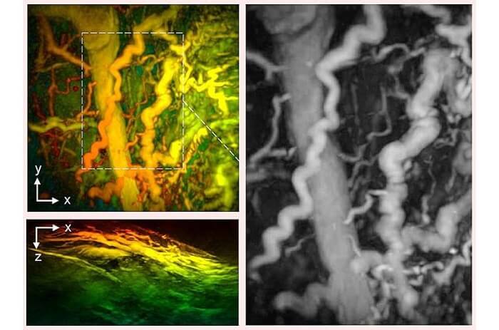
In pre-clinical tests, the device was used on 10 patients with conditions like diabetes, rheumatoid arthritis, and breast cancer. The scanner successfully produced detailed images of blood vessels, highlighting deformities and inflammation. For diabetes patients, the device revealed structural changes in foot blood vessels, offering insights into potential complications.
Associate Professor Andrew Plumb from UCL emphasized the device’s potential to monitor and understand disease progression, noting that it could provide vital information for early diagnosis.
The researchers plan to conduct further studies with larger patient groups to confirm the scanner’s clinical utility. The technology could become a valuable tool in diagnosing various conditions within the next three to five years, subject to further testing.