Researchers at Saarland University, led by Professor Bergita Ganse, have introduced a groundbreaking method to monitor bone healing without the use of harmful radiation. By focusing on blood flow and oxygen levels at the fracture site, their approach leverages near-infrared light—offering a safe, quick, and non-invasive alternative to traditional imaging techniques.
Moving Beyond X-rays and CT Scans
Until now, physicians have relied heavily on X-rays and CT scans to assess bone healing. These tools, while effective, expose patients to high-energy radiation and only provide occasional “snapshots” of the healing process. This can leave early signs of complications undetected.
Ganse and her team aim to fill that gap. In their recent publications in Biosensors and Bioelectronics and the Journal of Functional Biomaterials, they describe how standard blood flow and oxygen saturation devices—commonly used in skin and muscle diagnostics—can be adapted for bone monitoring.
Lighting the Way with Near-Infrared Technology
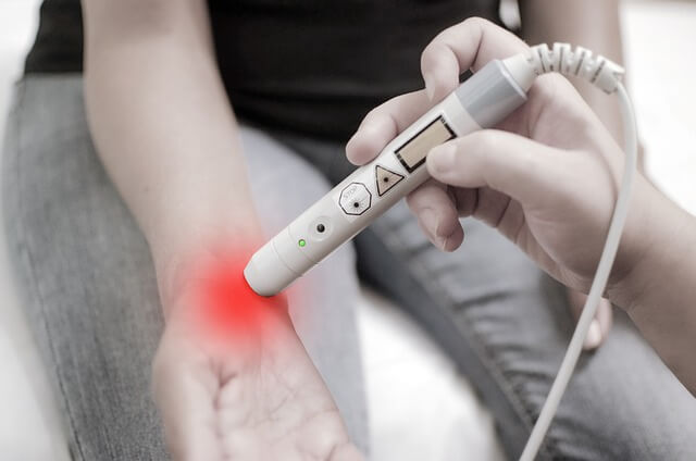
“These commercially available devices use harmless LED and laser light that penetrate deep enough to reach bone tissue,” explains Ganse. Her team successfully applied this technique in patients with fractured tibias, measuring healing progress through the skin—even through a plaster cast with a small opening.
As per Medical Xpress, this development could become a standard component of post-operative care worldwide.
Supplementing, Not Replacing, Imaging
Ganse emphasizes that their technique is not a substitute for X-ray imaging but a complementary tool. “It’s a rapid control method that fills in the blanks between standard scans,” she says. By tracking changes in oxygen and blood supply continuously, clinicians can detect complications early and respond promptly.
Traditional imaging often misses early healing because the soft bone tissue isn’t dense enough to show up. CT and X-ray images only reveal mineralization—when calcium salts strengthen the bone—which occurs later.
Real-Time Healing Insights
In contrast, the Saarland team’s method allows for continuous, real-time monitoring. Patients gain a clearer understanding of their recovery progress, and physicians can intervene early if healing stalls.
“Out of every 100 lower leg fractures, 14 develop complications. These are often detected too late,” says Ganse. With earlier detection, interventions like pulsed ultrasound, shockwave therapy, or magnetic field therapy can significantly improve outcomes.
Practical and Scalable for Global Use
The technology’s simplicity and affordability make it especially valuable in resource-limited or remote areas. “We could see these small devices used anywhere—from modern hospitals to rural clinics,” Ganse notes.
Understanding the Science Behind Healing
Fracture repair unfolds in distinct phases. Initially, fibrous tissue bridges the fracture gap. Then, new bone forms and receives a fresh blood supply as vessels regenerate.
In studies involving 55 tibial fracture patients and 51 healthy controls, Ganse’s team charted the evolution of blood flow and oxygen saturation throughout healing. “We observed a clear pattern,” she says. Blood flow surged early, peaked within weeks, and then declined. Meanwhile, oxygen saturation dropped initially but rose again as new vessels formed.
Detecting Problems Before They Show Up on Scans
If the readings don’t normalize, it may signal trouble. The team uses a device that combines laser Doppler for blood flow and white light spectroscopy for oxygen levels. “This method may detect issues earlier than X-rays ever could,” says Ganse.
Delayed healing could stem from excessive movement, poor immobilization, or health conditions like smoking or cancer. “With conventional imaging, these red flags are often visible only at advanced stages,” Ganse adds.
Challenges and Future Directions
One current limitation is depth—light-based measurements can’t assess fractures deeper than five centimeters beneath the skin. Still, Ganse’s team is pushing boundaries through other innovations.
They’re developing self-sensing shape memory materials that track stiffness and elasticity changes as bones heal. This effort is part of the Smart Implants project, a multidisciplinary initiative combining expertise from medicine, engineering, and computer science.
Smart Implants: Healing and Monitoring in One
The team has already created prototype fracture plates that actively support healing. Some offer micromechanical stimulation or adapt their stiffness based on recovery progress. They’re also working to embed the light-based sensors into intramedullary nails—devices only millimeters wide.
Expanding the Reach of the Technology
Now, Ganse and her colleagues are adapting their method to monitor other types of fractures. “It’s exciting to see how quickly this could move from research into routine clinical care,” she says.
Her unique background in space medicine adds depth to her work. In collaboration with the European Space Agency (ESA), the German Aerospace Center (DLR), and NASA, Ganse studies musculoskeletal degeneration in astronauts. Her insights are now helping patients on Earth recover faster and safer from fractures.


