
















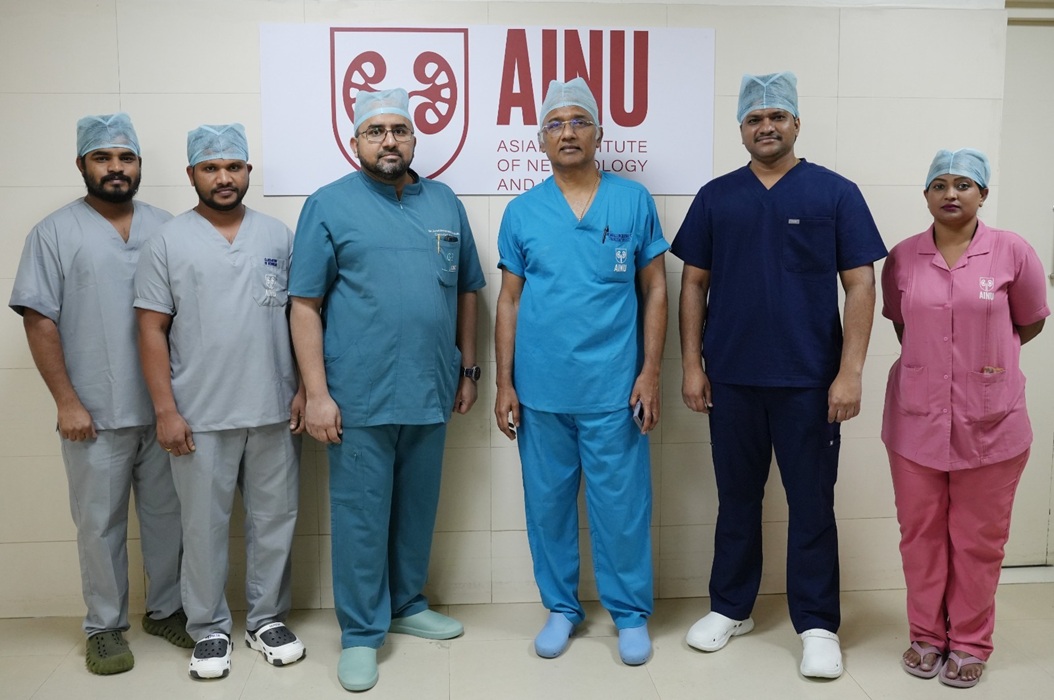
Doctors at the Asian Institute of Nephrology and Urology (AINU), Hyderabad, successfully treated a six-year-old boy diagnosed with a rare kidney tumour despite his having one of the rarest congenital kidney abnormalities—a crossed fused ectopic kidney. Using a highly specialised minimally invasive laparoscopic procedure, the surgical team removed the tumour while preserving healthy kidney tissue and function.
Rare Condition Presents Complex Surgical Challenge
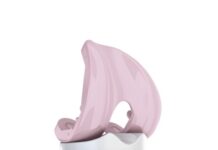
The child was brought to AINU after he experienced blood in his urine for two days. Detailed investigations revealed a tumour in a crossed fused ectopic kidney, a rare congenital condition in which both kidneys are fused together and positioned on the same side of the body.
This uncommon anatomical abnormality presents significant surgical challenges because the kidneys have distorted collecting systems and an unpredictable network of blood vessels. As a result, conventional surgical approaches carry a much higher risk of complications.
Advanced Imaging Enables Precision Surgery
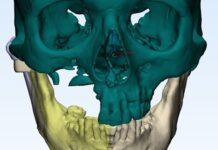
To address these challenges, the surgical team first created a customised surgical plan using advanced three-dimensional imaging to accurately map the child’s unique vascular anatomy. During the procedure, they also used laparoscopic intraoperative ultrasound to precisely identify the tumour’s location, depth and margins within the fused kidney.
As per the press release, these advanced imaging technologies enabled surgeons to selectively control only the blood vessels supplying the tumour while preserving blood flow to the healthy portions of the kidney. The team removed the tumour completely through tiny keyhole incisions and achieved clear surgical margins.
Following the surgery, the child experienced no major complications, retained normal kidney function and recovered well before being discharged in good health.
Expert Speaks
Commenting on the rare condition and the surgical procedure, Dr. Syed Md. Ghouse, Director – Robotics and Minimal Access Urology, AINU, who led the surgical team, said:
“Operating on a tumour in a crossed fused ectopic kidney is among the most demanding procedures in paediatric urology because every patient’s anatomy is unique and conventional surgical landmarks are absent. Success in such cases depends on meticulous planning and the intelligent use of advanced technologies. Three-dimensional reconstruction, intraoperative ultrasound and ICG fluorescence imaging allowed us to precisely identify the tumour and preserve the healthy kidney, giving the child the best possible long-term outcome while avoiding the trauma of open surgery.”
Preserving Kidney Function Was the Priority
Speaking about the procedure, Dr. Mallikarjuna C, Managing Director, Chief Consultant Urologist & Robotic Surgeon, AINU, said:
“Managing tumours in a fused kidney is exceptionally complex due to the abnormal anatomy and shared blood supply. Using advanced keyhole surgical techniques, we were able to precisely remove the tumour while preserving maximum kidney function—something that is critical, especially in such rare cases.”